Diet-Associated Dilated Cardiomyopathy in Dogs: From Grain-Free Diets to Cellular Biology — Part 3
Diet-Associated Dilated Cardiomyopathy in Dogs: From Grain-Free Diets to Cellular Biology — Part 3

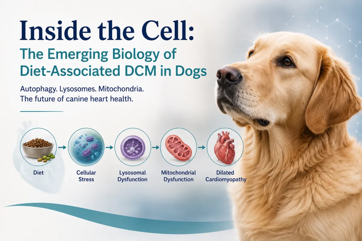
Inside the Cell: Autophagy, Lysosomes, Phospholipidosis, and the Future of Diet-Associated DCM
By Christina Bové, DVM, MS, DACVIM (Cardiology)
In Parts 1 and 2, we followed the evolution of diet-associated dilated cardiomyopathy from early reports of taurine deficiency to modern metabolomic studies that demonstrated complex alterations in nutrient metabolism.
Those discoveries answered an important question:
Diet appears capable of altering cardiac physiology.
But they created an even bigger one.
What is actually happening inside the heart cell?
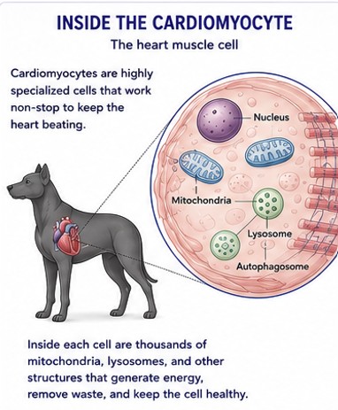
Today, researchers are looking beyond nutrients, ingredients, and even metabolism itself. The newest studies are focused on the cardiomyocyte—the individual heart muscle cell—and the intricate systems responsible for maintaining its health.
Instead of asking which ingredient might be responsible, investigators are exploring how diet may influence cellular recycling, energy production, lipid metabolism, and organelle function.
Although much of this work is still emerging, it represents one of the most exciting developments in veterinary cardiology.
The Heart Is Constantly Rebuilding Itself
Every heartbeat requires an enormous amount of energy.
Unlike skeletal muscle, which contracts only when needed, the heart contracts continuously throughout life without resting. To sustain this remarkable workload, cardiomyocytes must constantly repair themselves, replace damaged components, and generate enough energy for the next contraction.
This means the heart isn't simply pumping blood—it is continuously rebuilding itself.
Every heart muscle cell must:
Replace damaged proteins
Recycle worn-out mitochondria
Remove oxidized lipids
Maintain healthy cellular membranes
Prevent the accumulation of toxic cellular waste
When these protective systems function efficiently, the heart continues to perform despite decades of continuous work.
When they begin to fail, cellular stress gradually accumulates.
Autophagy: The Cell's Recycling Program
One of the most important protective mechanisms inside every cardiomyocyte is autophagy.
The word literally means "self-eating," but the name is somewhat misleading.
Rather than destroying healthy tissue, autophagy is one of the body's most important housekeeping systems.
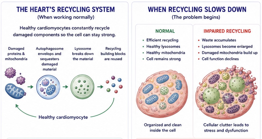
Damaged proteins, dysfunctional mitochondria, and worn-out cellular components are enclosed within specialized membrane-bound structures called autophagosomes. These then fuse with lysosomes, where their contents are broken down into reusable building blocks.
The cell doesn't simply dispose of damaged material—it recycles it.
Without this continual renewal process, damaged proteins, abnormal lipids, and dysfunctional organelles would gradually accumulate inside the cardiomyocyte, reducing efficiency and increasing cellular stress.
For a heart that beats more than 100,000 times every day, efficient cellular recycling is essential.
When Cellular Recycling Slows Down
Researchers are increasingly investigating whether alterations in cellular recycling contribute to diet-associated DCM.
Although the precise mechanisms remain under investigation, recent experimental studies suggest that diets associated with DCM may influence metabolic pathways involved in maintaining normal cellular health.
Rather than causing immediate structural heart disease, these metabolic changes may gradually impair the heart cell's ability to repair itself.
If damaged proteins, lipids, and organelles are not efficiently removed, cellular stress may begin to accumulate long before clinical heart disease becomes apparent.
This represents an important shift in thinking.
Instead of asking whether a single nutrient is deficient, investigators are now exploring whether diet alters the systems responsible for maintaining healthy cardiomyocytes.
Lysosomes: More Than the Cell's Garbage Disposal
At the center of this recycling system are lysosomes.
Often described as the cell's garbage disposal, lysosomes are actually far more sophisticated.
They play critical roles in:
Autophagy
Lipid metabolism
Nutrient sensing
Intracellular signaling
Mitochondrial quality control
When lysosomes function normally, damaged proteins, phospholipids, and dysfunctional organelles are efficiently broken down and recycled.
When lysosomal function becomes impaired, however, cellular waste begins to accumulate.
Over time, this accumulation may interfere with normal myocardial function.
Phospholipidosis: A New Piece of the Puzzle
As researchers shifted from studying nutrients to studying cells, another intriguing finding began to emerge: phospholipidosis.
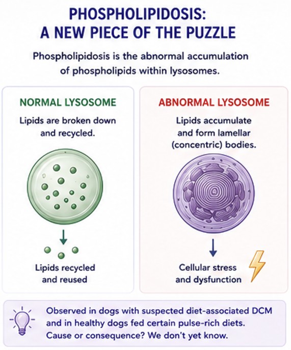
Although the name sounds intimidating, phospholipidosis simply describes the abnormal accumulation of phospholipids—essential components of cell membranes—within intracellular structures called lysosomes.
To understand why this matters, it helps to first understand what lysosomes do.
Think of lysosomes as the cell's recycling centers. They break down damaged proteins, worn-out organelles, and old cell membrane components into smaller building blocks that can be reused. This recycling system is essential for maintaining healthy cardiomyocytes, which must contract continuously throughout a dog's lifetime.
Normally, phospholipids are constantly broken down and replaced as cell membranes are repaired and renewed. However, if this recycling process becomes disrupted, phospholipids begin to accumulate within lysosomes, forming the characteristic microscopic lamellar bodies seen with phospholipidosis.
An Unexpected Discovery
One of the most intriguing recent findings came from work by Dr. Lisa Freeman and colleagues at Tufts University.
Using advanced microscopy and biochemical techniques, investigators identified phospholipid accumulation within the hearts of dogs with suspected diet-associated DCM. Similar changes were also observed experimentally after healthy dogs consumed certain pulse-rich diets.
These observations suggested that affected cardiomyocytes were experiencing abnormalities in intracellular lipid handling and cellular recycling.
Importantly, these findings moved the conversation beyond traditional nutrition.
Researchers were no longer asking whether dogs were deficient in a particular vitamin or amino acid.
Instead, they began asking whether certain dietary formulations might alter how heart muscle cells process, recycle, and maintain their internal components.
Cause or Consequence?
This is where the science becomes particularly interesting.
At present, researchers do not know whether phospholipidosis directly causes myocardial dysfunction or whether it develops secondary to other metabolic changes occurring within the heart.
In other words, phospholipidosis may represent:
A primary contributor to disease
A marker of impaired lysosomal function
OR
Simply evidence that cardiomyocytes are under metabolic stress
Each possibility has different implications, and ongoing studies are working to determine which is correct.
Why Researchers Are Excited
Although phospholipidosis has not been proven to cause diet-associated DCM, it provides one of the first tangible cellular abnormalities that helps connect dietary formulation with changes occurring inside the myocardium.
Rather than focusing on a single missing nutrient, investigators are beginning to view diet-associated DCM as a disorder involving multiple interconnected pathways, including nutrient metabolism, lipid trafficking, cellular recycling, and energy production.
This systems-based approach is remarkably different from where the field stood just a decade ago.
Lessons from Human Medicine
Interestingly, phospholipidosis is not unique to veterinary medicine.
In humans, similar intracellular phospholipid accumulation has been described with certain medications—such as amiodarone and chloroquine—as well as several inherited lysosomal storage disorders. In these conditions, disrupted lysosomal function can impair normal cellular homeostasis and contribute to tissue dysfunction.
Whether diet-associated DCM shares aspects of these mechanisms remains unknown. However, these parallels provide biologic plausibility for continued investigation and suggest that lysosomal dysfunction may represent an important area of future research.
What This Means Today
For practicing veterinarians, phospholipidosis should not change clinical recommendations at this time.
Instead, it changes how we think about the disease.
Rather than viewing diet-associated DCM as a simple taurine deficiency, the emerging evidence suggests it may represent a far more complex disorder involving altered metabolism, intracellular lipid handling, impaired cellular recycling, and mitochondrial health.
As Dr. Freeman and colleagues have emphasized, these findings are hypothesis-generating, not definitive proof of causation. Nevertheless, they provide an exciting glimpse into where the science is headed and may ultimately help explain why some dogs develop diet-associated DCM while millions of others eating similar diets remain completely healthy.
Mitochondria: Powering Every Heartbeat
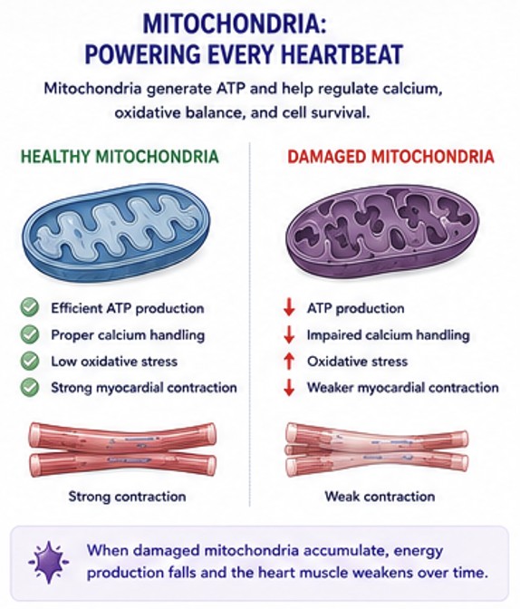
Ultimately, every pathway we've discussed converges on one critical objective: maintaining energy production within the heart muscle cell.
Healthy cardiomyocytes contain thousands of mitochondria that generate adenosine triphosphate (ATP), the energy required for every heartbeat. In fact, mitochondria occupy nearly one-third of the volume of a normal cardiomyocyte, underscoring just how energy-dependent the myocardium truly is.
Beyond producing ATP, mitochondria regulate calcium handling, oxidative balance, and programmed cell survival. Because the heart contracts continuously throughout life, maintaining a healthy population of mitochondria is essential for normal cardiac function.
This is where cellular recycling becomes critically important.
Damaged mitochondria are normally identified and removed through a specialized form of autophagy known as mitophagy. When this quality-control system functions properly, dysfunctional mitochondria are replaced before they can impair cellular performance.
However, if cellular recycling slows—or lysosomal function becomes impaired—damaged mitochondria may accumulate. The result is reduced ATP production, increased oxidative stress, and diminished contractile efficiency.
Less energy means weaker contraction.
Over time, this may contribute to impaired systolic function, ventricular remodeling, and eventually the clinical phenotype of dilated cardiomyopathy.
Although much remains to be learned, this emerging model provides a biologically plausible explanation for how dietary alterations could influence cardiac performance without causing a traditional nutrient deficiency.
Putting the Pieces Together
Over the past decade, researchers have moved from studying nutrients, to studying metabolic pathways, and now to studying the biology of the cardiomyocyte itself.
Rather than identifying a single missing nutrient, investigators are beginning to recognize that diet-associated DCM may result from disruption of multiple interconnected cellular processes.
While no single mechanism has been proven, a working hypothesis is beginning to emerge:
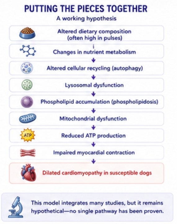
This model remains hypothetical, but it integrates many of the observations reported over the past decade—including metabolomic studies, foodomics research, feeding trials, ultrastructural findings, and emerging cellular biology. Importantly, it also helps explain why diet-associated DCM behaves differently from inherited DCM and why many affected dogs experience meaningful reverse remodeling following dietary modification.
Why Do Only Some Dogs Become Affected?
One of the most important questions facing researchers today is no longer whether diet can contribute to dilated cardiomyopathy. The cumulative evidence—including reverse remodeling after dietary modification, metabolomic studies, feeding trials, and emerging cellular research—strongly supports a diet-associated form of DCM in susceptible dogs.
The question has now become:
Why do some dogs develop disease while others consuming similar diets do not?
This question has fundamentally changed the direction of research.
If dietary formulation were the only factor, clinicians would expect a far more predictable pattern of disease. Conversely, if genetics alone explained the condition, affected dogs would not consistently improve following dietary modification, and controlled feeding studies would not demonstrate measurable metabolic and cardiac changes.
Current evidence suggests that diet serves as the initiating factor, while host-specific factors influence an individual dog's response.
Researchers are actively investigating several contributors to susceptibility, including:
Genetics. Individual variation in amino acid metabolism, lipid handling, mitochondrial function, and cellular stress responses.
Dietary formulation. Pulse burden, pulse diversity, ingredient processing, and nutrient interactions may all influence risk.
Nutrient bioavailability. Two diets may contain similar nutrient concentrations on paper but differ substantially in digestibility and metabolic availability.
Gut microbiome. Intestinal bacteria influence amino acid metabolism, bile acid recycling, inflammation, and numerous metabolic pathways that may affect cardiac health.
Individual metabolism. Dogs likely differ in their ability to synthesize taurine, regulate lipid metabolism, maintain mitochondrial function, and adapt to dietary challenges.
Concurrent disease. Gastrointestinal disease, endocrine disorders, obesity, age, and other medical conditions may alter nutrient metabolism and increase susceptibility.
Rather than acting independently, these factors likely interact.
A particular dietary formulation may be well tolerated by one dog but create sufficient metabolic stress in another to initiate the cascade described throughout this series—from altered nutrient metabolism and impaired cellular recycling to lysosomal dysfunction, phospholipid accumulation, mitochondrial dysfunction, and ultimately impaired myocardial contraction.
The next frontier of research is no longer proving that nutrition matters. Instead, investigators are working to identify which dogs are most susceptible, why they are susceptible, and how we can recognize those patients before irreversible myocardial injury develops.
Answering those questions could fundamentally change canine nutrition by allowing veterinarians to identify at-risk patients, personalize dietary recommendations, and intervene before structural heart disease develops.
What Does This Mean for Veterinarians?
Although the cellular mechanisms continue to evolve, the practical approach to suspected diet-associated DCM remains remarkably consistent.
Veterinarians should continue to:
Obtain a complete diet history in every dog diagnosed with DCM
Identify diets with a potentially high pulse burden
Measure whole blood and plasma taurine concentrations when appropriate
Perform a comprehensive cardiac evaluation, including echocardiography when DCM is suspected
Recommend dietary modification when diet-associated DCM is suspected
Recognize that many affected dogs can experience meaningful reverse remodeling following intervention.
Importantly, the absence of a fully defined mechanism should not delay appropriate diagnosis or treatment. Clinical decisions should continue to be guided by the best available evidence while research continues to refine our understanding.
Key Takeaways
The understanding of diet-associated dilated cardiomyopathy has evolved dramatically over the past decade—from focusing on individual nutrients to investigating complex cellular biology.
Emerging evidence suggests that altered metabolism, impaired cellular recycling, lysosomal dysfunction, phospholipid accumulation, and mitochondrial health may all contribute to disease development.
Phospholipidosis has emerged as one of the most intriguing recent discoveries, but it remains an observation rather than a proven cause of myocardial dysfunction.
Diet-associated DCM is likely a multifactorial disease resulting from the interaction between dietary formulation and individual susceptibility.
The field has shifted from asking "What nutrient is missing?" to "How does diet influence the biology of the cardiomyocyte?"
Looking Ahead
Over the past three articles, we've followed one of the most fascinating scientific journeys in modern veterinary cardiology.
We began by examining the early concerns surrounding grain-free diets and the recognition that certain dietary formulations were associated with a potentially reversible form of dilated cardiomyopathy. We then explored the history of taurine-responsive cardiomyopathy, the emergence of metabolomics and foodomics, and the landmark feeding studies that demonstrated measurable metabolic and cardiac changes in healthy dogs.
In this article, we moved one step deeper—inside the cardiomyocyte itself.
Although many questions remain unanswered, one message has become increasingly clear:
Diet-associated DCM is unlikely to be explained by a single ingredient, nutrient, or metabolic pathway.
Instead, the disease appears to involve a complex interaction between dietary formulation, metabolism, cellular recycling, lipid handling, mitochondrial health, and the individual dog's biologic response.
This represents a remarkable shift in our understanding.
The conversation has progressed from ingredients...
...to nutrients...
...to metabolism...
...and now to the biology of the heart cell itself.
Perhaps that is the most important lesson of all.
Nutrition is not simply about meeting minimum nutrient requirements. Every meal influences metabolism, every metabolic pathway influences cellular function, and every healthy cardiomyocyte depends on those systems working together to generate the energy required for a lifetime of heartbeats.
As researchers continue to investigate these mechanisms, our understanding of diet-associated DCM will undoubtedly continue to evolve.
But while the science has become increasingly sophisticated, veterinarians and dog owners are still faced with practical questions every day.
Which diets should we recommend?
Should healthy dogs avoid pulse-rich diets?
How should veterinarians evaluate ingredient lists?
What does the current evidence actually support?
Those are the questions we'll answer in Part 4, where we'll move from the laboratory back to the clinic and translate the science into practical, evidence-based recommendations for veterinarians and dog owners.
Because ultimately, understanding the biology is only the first step.
Applying it to improve the health of our patients is what matters most.
References
Freeman LM, Rush JE, Berridge BR, Mitchell RN, Martinez-Romero EG, et al. Dogs with diet-associated dilated cardiomyopathy have higher urine di-docosahexaenoyl-bis(monoacylglycerol)phosphate, a biomarker of phospholipidosis. American Journal of Veterinary Research. 2025.
Smith CE, Freeman LM, Rush JE, et al. Foodomics Analysis of Commercial Dog Foods Associated with Dilated Cardiomyopathy. Scientific Reports. 2021.
Smith CE, Freeman LM, Rush JE, et al. Metabolomic Profiling of Dogs with Suspected Diet-Associated Dilated Cardiomyopathy. Scientific Reports. 2022.
Quilliam CK, et al. Effects of Pulse-Based Diets on Nutrient Digestibility and Metabolic Responses in Healthy Dogs. Frontiers in Veterinary Science. 2021.
Quilliam CK, et al. Effects of a 28-Day Feeding Trial of Grain-Containing Versus Pulse-Rich Diets on Cardiac Function, Taurine Status, and Nutrient Digestibility in Healthy Dogs. PLOS ONE. 2023.
Freeman LM, Stern JA, Fries R, Adin DB, Rush JE. Diet-Associated Dilated Cardiomyopathy in Dogs: What Do We Know? Journal of the American Veterinary Medical Association. 2018.
Adin DB, et al. Clinical Findings and Outcome of Dogs with Suspected Diet-Associated Dilated Cardiomyopathy. Journal of Veterinary Internal Medicine. 2021.
Walker AL, et al. Clinical and Echocardiographic Features of Dogs with Suspected Diet-Associated Dilated Cardiomyopathy. Journal of Veterinary Cardiology. 2021.
Kaplan JL, Stern JA, Fascetti AJ, et al. Taurine Deficiency and Dilated Cardiomyopathy in Golden Retrievers Fed Commercial Diets. PLOS ONE. 2018.
Ontiveros ES, et al. Development of Taurine Deficiency and Dilated Cardiomyopathy in Golden Retrievers Fed Commercial Diets. PLOS ONE. 2020.
Klionsky DJ, et al. Guidelines for the Use and Interpretation of Assays for Monitoring Autophagy. Autophagy.
Ballabio A, Bonifacino JS. Lysosomes as Dynamic Regulators of Cell and Organismal Homeostasis. Nature Reviews Molecular Cell Biology. 2020.
Mizushima N, Komatsu M. Autophagy: Renovation of Cells and Tissues. Cell. 2011.
Schulze H, Sandhoff K. Lysosomal Lipid Storage Diseases. Cold Spring Harbor Perspectives in Biology. 2011.
Matsui Y, Takagi H, et al. Molecular Mechanisms and Pathophysiological Significance of Autophagy During Myocardial Ischemia and Heart Failure. Autophagy.
Dorn GW II. Mitochondrial Dynamics in Heart Disease. Biochimica et Biophysica Acta (BBA) – Molecular Cell Research.
Dorn GW II, Vega RB, Kelly DP. Mitochondrial Biogenesis and Dynamics in the Developing and Diseased Heart. Genes & Development. 2015.
World Small Animal Veterinary Association (WSAVA) Global Nutrition Committee. Global Nutrition Toolkit.
U.S. Food and Drug Administration. Investigation into Potential Link Between Certain Diets and Canine Dilated Cardiomyopathy. FDA Updates (2018–present).
Freeman LM. Tufts Petfoodology: Diet-Associated Dilated Cardiomyopathy Resource Articles. Cummings School of Veterinary Medicine at Tufts University.
